by Dr. Yuhong Dong and Dr. Ann Corson (Epoch Times)

Fibrous Clots, Foreign Matter in Blood After COVID Jabs: Is There a Way to Detox?
Recently unusual blood clots as well as metal-like foreign objects found in the vessels of COVID-19 jab recipients have been reported across the country. Both types of substances are unusual and are likely to be harmful to our bodies. What are the potential causes and ramifications of these substances, and is there any chance of reversing the mysterious condition?
The Korea Veritas Doctors (KoVeDocs) for COVID-19 previously found certain foreign materials and moving parasite-like entities in the Pfizer and Moderna mRNA COVID-19 vaccines as those vaccines were warmed to near room temperature.
Subsequently, on March 11 2022, three Korean doctors, Young Mi Lee, Sunyoung Park, and Ki-Yeob Jeon, published findings of similar foreign materials in samples of blood from COVID-19 jab recipients in a paper titled “Foreign Materials in Blood Samples of Recipients of COVID-19 Vaccines” in the International Journal of Vaccine Theory, Practice, and Research.
The various shapes and sizes of the foreign materials in centrifuged plasmas of COVID-19 vaccinated individuals closely resembled the shapes and sizes of foreign materials previously observed directly in the vaccines themselves.
The evidence suggests that the foreign materials found in the COVID-19 vaccine recipients in this study were injected into their bodies when they received one or more doses of the COVID-19 vaccines.
The Italian doctors have repeated the Korean study in a much larger sample and with a more advanced technique, i.e. dark-field microscopic analysis of fresh peripheral blood on a slide, allowing a first and immediate assessment of the health status of a person’s blood, better representing their overall health status.
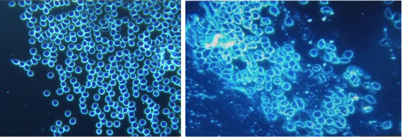
One month after the mRNA inoculation, a total of 948 subjects (94 percent of the total studied population) showed blood with aggregation of erythrocytes (red blood cells) and the presence of particles of various shapes and sizes of unclear origin.
This foreign material seemed to collect itself into structures, sometimes forming crystals and other times forming long tubes or fibers.
The foreign structures in the patients’ blood, which had not been there before vaccination, certainly look unusual in the photos included in the study.
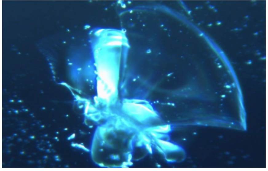
This image at 120x magnification (3x magnification digitally produced) hihglights a typical self-aggregating structure in fibro/tubular mode. (Courtesy of IJVTPR)
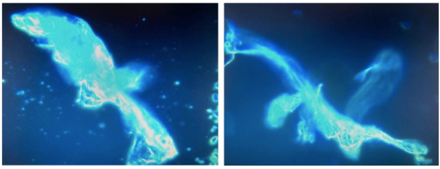
These photos are at 40x magnification. At the left side, (a) shows the blood condition of the patient before the inoculation. The right side image, (b) shows the same person’s blood one month after the first dose of Pfizer mRNA “vaccine.” Particles can be seen among the red blood cells which are strongly conglobated around the exogenous particles; the agglomeration is believed to reflect a reduction in zeta potential adversely affecting the normal colloidal distribution of erythrocytes as see at the left. The red blood cells at the right (b) are no longer spherical and are clumping as in coagulation and clotting. (Courtesy of IJVTPR)
Twelve subjects, whose blood was examined with the same method before vaccination, showed a perfectly normal blood appearance under the microscope. The alterations found after the inoculation of the mRNA injections further reinforce the suspicion that the changes were due to the jabs themselves.
These photos are at 40x magnification. At the left side, (a) shows the blood condition of the patient before the inoculation. The right side image, (b) shows the same person’s blood one month after the first dose of Pfizer mRNA “vaccine.” Particles can be seen among the red blood cells which are strongly conglobated around the exogenous particles; the agglomeration is believed to reflect a reduction in zeta potential adversely affecting the normal colloidal distribution of erythrocytes as seen at the left. The red blood cells at the right (b) are no longer spherical and are clumping as in coagulation and clotting. (Courtesy of IJVTPR)
In August 2022, the Italian doctors’ results were published in the same journal as the Korean data.
A German Working Group for COVID Vaccine Analysis, an interdisciplinary working group that undertook the task of analyzing the contents and effects of the novel COVID-19 vaccines, also examined the results.The aforementioned group consists of independent scientists, including doctors, physicists, chemists, microbiologists, pharmacologists, and alternative health practitioners, supported by lawyers, psychologists, analysts and journalists.
In July 2022, they published their preliminary findings which are quite similar to the findings of the aforementioned studies.
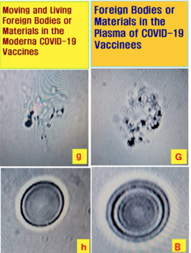
They found visible distinctive particles with complex metallic structures of different sizes under the dark-field microscope in the blood of all 48 vaccinated people. Those particles were like crystalline formations. Without exception, all of these patients showed peculiarities that were not observed in a single case of unvaccinated subjects.
They have established that the COVID-19 vaccines consistently contain substances with compositions that cannot be determined. Some ingredients have not even been listed as ingredients by the vaccine manufacturers.Findings From Embalmers: Numerous Long, String-like Fibrous Clots
Several embalmers across the country
have been observing many large, and sometimes very long, “fibrous” and
“rubbery” clots inside corpses, starting from either 2020 or 2021.
Mike Adams, who runs an ISO-17025 accredited lab in Texas, analyzed clots in August and found them to be lacking iron, potassium, magnesium, and zinc.
The string-like structures differ in size, but the longest can be as long as a human leg and the thickest can be as thick as a pinky finger.
Richard Hirschman, a licensed funeral director and embalmer in Alabama, said “Prior to 2020, 2021, we probably would see somewhere between 5 to 10 percent of the bodies that we would embalm [having] blood clots,” however now, 50 percent to 70 percent of the bodies he sees have clots.
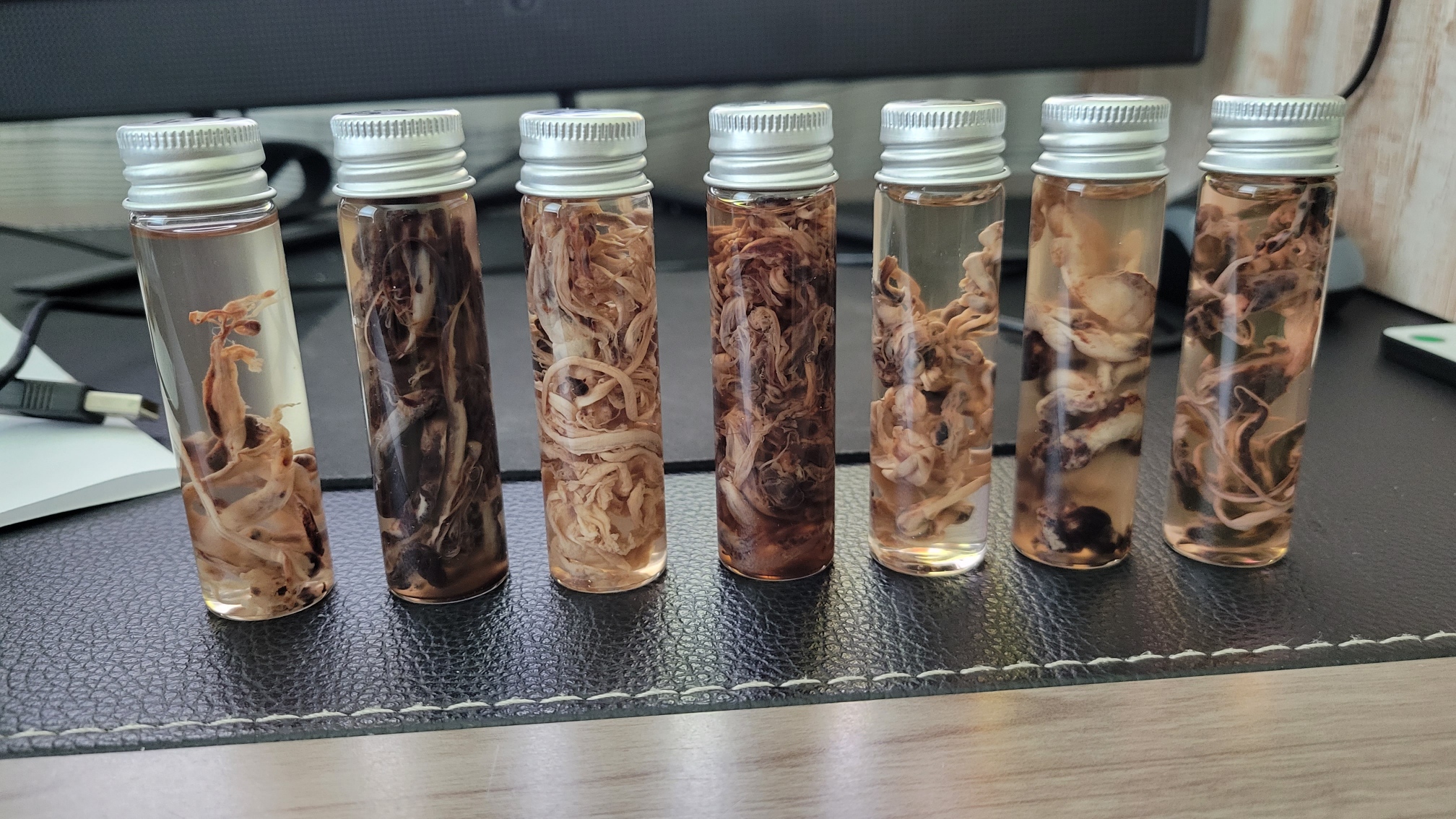
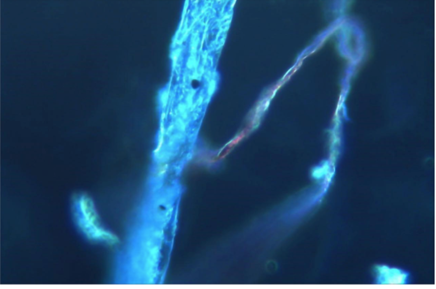
Fibrous Clots found in corpses by Richard Hirschman (Courtesy of Richard Hirschman)
Where Do They Come From?
Everyone wants to know what exactly is the cause of these strange, fibrous clots in the blood. But answers may be hard to find, for the manufacturers will not easily disclose the crux of mRNA vaccines.
What we do know is that there are two main components that form these strange things in the blood: metallic elements and protein components. But where do they come from?
Metallic Materials
- What is graphene?
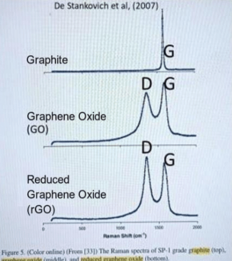
Graphene oxide (GO) is derived from graphite by oxidation treatment, which retains the large planar structure and high surface area while introducing hydrophilic groups such as hydroxyl, carboxyl, and epoxy groups. It so becomes a potential carrier platform for bioactive molecules because of unique physicochemical properties, especially the surface that can be easily modified.
GO nanomaterials have been widely used in biomedical fields due to their unique properties such as drug delivery, biological sensor, photodynamic therapy, cancer therapy, antibacterial therapy, and vaccines.
GO has been reported to be used as the delivery vehicle of vaccine antigen, which delivers antigen into dendritic cells (DCs: professional cells reporting signals to T cells). Antigen-loaded GO promotes the cross-presentation of antigen to CD8+T cells, which contributes to the elimination of intracellular pathogens and pathogenic cells after vaccination.
A peer reviewed article has discussed the use of graphene as an adjuvant in vaccines.
On the other hand, an in vitro study showed that GO can activate the immune system by increasing inflammatory factors as well as the proliferation and differentiation of lymphocytes, especially CD8+T cells.
Although GO shows enormous potential in the preparation of new vaccines, its low solubility and poor stability limits its application in the human body.
Furthermore, GO has been shown to be cytotoxic, which may be related to the proteins on its surface; especially since it can damage DNA, a property associated with its inherent chemical characteristics.
GO has been used in flu shots. There is a possibility of graphene oxide being used in the vaccines of COVID for the purpose of increasing the immunogenicity, i.e. the ability of a vaccine to induce an immune response, which is often evaluated by the concentration of neutralizing antibodies in vaccine development trials.
- Identification of graphene in COVID Vaccines
La Quinta Columna, is a Spanish research team founded by Ricardo Delgado and Dr. José Luis Sevillano.
Dr. Pablo Campra Madrid, PhD in Chemical Sciences and Bachelor in Biological Sciences, associate professor at the University of Almeria, Spain was the first to examine the COVID jabs for metallic materials. He did this at the request of La Quinta Columna.
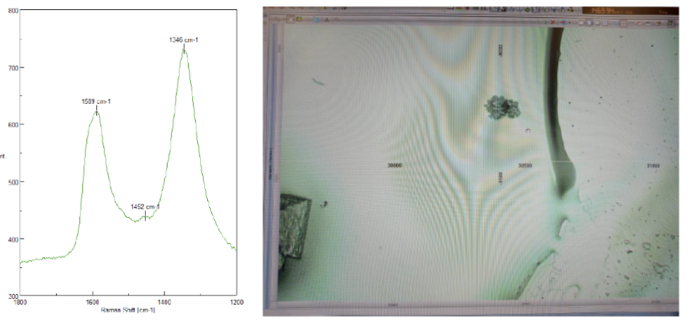
Dr. Campra Madrid published a report in November 2021 entitled “Detection of Graphene in COVID19 vaccines by micro-raman spectroscopy.” (An English translation of his report can be found here).
He carried out a screening of nanoparticles visible with the optical microscopy in seven random samples of vials of COVID vaccines from four different manufacturers.
Using a technique called micro-RAMAN, Dr. Campra was able to determine the presence of graphene in the samples.
RAMAN infrared spectroscopy is a fast technique that allows us to detect the structure of a material without altering or destroying its properties. It is a modern technique named after its developer Sir C. V. Raman.
Coupled optical microscopy allows the excitation laser to be focused on specific objects and points located on objects, to reinforce the degree of confidence in identifying the nature of the material, and to obtain complementary information on thickness, defects, thermal conductivity and edge geometry of graphene nanocrystalline structures.
(“Detection of Graphene in COVID19 vaccines by micro-raman spectroscopy.”)
After screening, more than 110 objects were selected for their graphene-like appearance under optical microscopy. Of these, a group of 28 objects were selected due to the compatibility of both images and spectra with the presence of graphene derivatives, based on the correspondence of these signals with those obtained from standards and scientific literature.
The identification of graphene oxide structures can be regarded as conclusive in 8 out of 28 due to the high spectral correlation with the standard. In the remaining 20 objects, images coupled with Raman signals show a very high level of compatibility with undetermined graphene structures, despite the different methods used for detection. .
Here is one sample of the Pfizer vaccine (code: Pfizer 2 WBR UP GO2). The left side photo is the RAMAN signal which is very similar to the Graphene Oxide as above; the right photo shows the microscopic image of metallic aggregates (at a magnification of 100X).
As a conclusion of his report, which he has made freely available, Dr. Campra makes a call for ongoing studies, discussion, and replication of his work. He also asks other independent researchers, with no conflict of interest or coaction from any institution, to make wider counter-analysis of these products to achieve a more detailed knowledge of the composition and potential health risk of these experimental drugs.
Public Documentations of mRNA Vaccines
However, neither Pfizer nor Moderna’s public documentation claim their products contain graphene oxide or any other type of metallic components. Nevertheless, there are still potential incidences of quality issues during their mRNA’s manufacturing process, which have been reported from time to time since September 2021.
The issue of mRNA stability is a historically well-known issue in biological research. Unsurprisingly, the quality issues would frequently come up in the pharmaceutical industry, especially for an unstable component such as mRNA.
In the late summer of 2021, metallic contaminants were found in Moderna vaccine vials in Japan. As a result, Japanese authorities suspended the use of three Moderna batches consisting of 1.63 million doses.
Two men aged between 30 and 40 died within days of receiving the Moderna COVID-19 vaccine from the batches in question.
Also, a few weeks later, white floating matter was found in two unused vials of the Pfizer COVID-19 vaccine. Recently, Moderna had to recall 764,900 doses of its COVID-19 vaccine in Europe after contamination was found.
Campra reminds readers that graphene materials are potentially toxic to human beings and the presence of graphene in the COVID-19 vaccines has not been declared in any of the emergency use authorizations.
Proteins
Where do these fibrous proteins come from and how do they form insoluble fiber-like components?
It has been found that these clots are lacking key elements present in healthy human blood, such as iron, potassium, and magnesium, suggesting that they are formed from something other than blood.
The COVID vaccines, after inoculation, instruct the cells to produce large quantities of spike proteins. Normal biochemical and physiological processes are “hijacked” in order to make an abnormal amount of these spike proteins.
Spike proteins are able to form amyloid-like substances, unfold and form a different configuration, contributing to tight string-like bonded structures with longitudinal twisting as well as cross binding- such a process can be made visible through microscopy.
(“Amyloidogenesis of SARS-CoV-2 Spike Protein” in JACS)
Furthermore, the spike protein can cause blood clots by competitive binding to heparan sulfate; S1 (a part of the spike protein) can induce the production of fibrin resistant to fibrinolysis, leading to unopposed microclot formation.
Interaction Between Metallic Materials, Proteins and Human Body
Finally, there is an interaction between proteins, metals and the human body. It is estimated that approximately half of all our human cellular proteins can bind to metals (pdf).
A good example is the interaction between hemoglobin and iron (Fe). Hemoglobin is a protein found in red blood cells. The main function of hemoglobin is to carry oxygen throughout our body. It also transports some amount of carbon dioxide from different parts of the body to the lungs.
Hemoglobin is made up of four polypeptide subunits, two alpha (α) subunits and two beta (β) subunits. Each of the four subunits contains a heme molecule that contains an iron atom.
Bad examples of interactions between proteins and metals occur with the Tau protein (an abnormal misfolded protein found in Alzheimer’s disease) and metallic elements in the pathogenesis of Alzheimer’s disease.
It has been confirmed that close interactions of the three metal ions Fe2/3+, Cu2+, and Zn2+ with the human tau protein can cause structural changes.
Transmission electron microscopy studies of the tau aggregates formed in the presence of metal ions suggest that the presence of metal ions influences the aggregation process. Fluorescence studies of full-length htau40 in the presence of Cu2+ indicate the formation of reactive oxygen species, which may contribute further to oxidative stress and neuron death.
If the metallic material in the blood of COVID vaccine recipients were possibly proven to be graphene oxide, there are a number of researches which have reported the impact of graphene oxide on biological systems with or without a potential interaction with electromagnetic fields (EMF).
In 2018 a group of Spanish scientists reported that graphene oxide nanosheets disrupt lipid composition, Ca2+ homeostasis and synaptic transmission in primary cortical neurons (pdf).
One Korea study published in 2016 in the journal of Advanced Healthcare Materials, showed that the combination of reduced GO (RGO) and pulsed EMFs enhances the neurogenic and adipogenic differentiation of human alveolar bone marrow stem cells (hABMSCs), synergically increases extracellular matrix (ECM) formation, membrane proteins, and metabolism. The paper also indicates that magnetic-field-irradiated RGO can induce magnetic moments, which then induces electric currents when the magnetic fields change.
Why is graphene or GO so hotly contended? It has many attractive physical properties, such as high conductivity, good transparency and nonlinearity. Graphene is also a promising material to construct reconfigurable miniaturized resonant antennas.
In the 2018 International Japan-Africa Conference on Electronics, Communications and Computations (JAC-ECC), Egyptian scientists (Zainud-Deen, Malhat, and Ghazi) reported that graphene has been used in telecommunication infrastructures.
Their full paper is titled “High Gain Graphene-Based Magneto-Electric Antenna for 5G Communications.”
Even though the interaction between graphene in the body and EMFs is not yet precisely understood, we do know that our living environment is full of various EMF fields emitted by cell phone towers, mobile phones, TVs, radios, electrical appliances, and microwave emitters. Graphene is a metal with excellent conductivity.
These EMFs may have an impact on our health as well. For example, a study published in the Journal of Clinical and Translational Research in October 2021 explored the effects of EMFs on the clinical severity of COVID-19 infections. The authors propose “a substantial overlap in pathobiology” between COVID-19 and wireless communications radiation exposure, especially 5G. They present evidence that clinical progression of COVID-19 could be generated by wireless communication radiation (WCR) exposure.
Throughout the summer of 2021, hundreds of amateur videos circulated on social media of people with metallic objects sticking to their bodies at or close to injection sites, indicating they had developed electromagnetic properties following vaccination. These observations were however quickly dismissed by the mainstream media and scientific community as an adhesive property of natural skin oils. Nevertheless, researchers from the European Forum for Vaccine Vigilance did a study on the electromagnetism of vaccinated persons in Luxembourg which found that magnets indeed adhered to vaccinated people’s skin.
Additionally, the Spanish research institute, La Quinta Columna has presented evidence that some vaccinated people not only present with magnetism but also that graphene toxicity can present with symptoms similar to COVID-19 illness.
A detailed analysis of the COVID-19 vaccines has also revealed the presence of nano routers, tubules, and circuits that may explain why some of those vaccinated emit bluetooth MAC addresses.
Spanish and French researchers have documented MAC addresses coming from the vaccinated but not the unvaccinated. Unfortunately, much of this research is being heavily censored and labeled as false by fact-checkers.
The interaction of graphene inside the human body with EMFs should be thoroughly investigated and the potential harm to human health should be diligently elucidated.
As we have witnessed an extraordinarily wide range of adverse reactions after COVID jabs, perhaps these metals and jelly-like strings in the blood are perhaps key reasons for sudden death, cardiac attack, fatigue, brain fog, or neuropsychological syndrome after vaccination.
Other adverse consequences that have been reported include:
autoimmune diseases, weakened immune systems, inflammatory conditions,
organ damage, high blood pressure, heart attacks, myocarditis,
neurodegenerative diseases, cancer, and shortened life expectancy.
A Global Collaboration Is Called to Further Investigation
As doctors who have been observing this pandemic and the vaccination agenda since the beginning, we have to express our deep concern in relation to these unprecedented findings.
All the scientists who have conducted their own research on this matter have expressed a warning and alerted the global medical community about the seriousness of this matter.
Nobody has ever witnessed such types of abnormal metals in addition to the unusual fibrotic string-like clots in humans.
The first recommendation would be to halt all COVID-19 vaccination programs immediately.
There should be an immediate cessation of and abandonment of any COVID-19 “Vaccine Pass Policy” and any other form of mandate for COVID-19 vaccinations.
People will likely ask what they should do if they have already been vaccinated, and how to determine if they have these components in their blood.
To answer this question, a collaborative worldwide evaluation of COVID-19 vaccine contents, and blood plasma samples of individuals vaccinated with the COVID-19 shots should be undertaken immediately with all due diligence.Emergency collaborative studies of detoxification protocols for COVID-19 vaccine sequelas should be undertaken.
How Should Vaccinated People Get Rid of These Foreign Metallic Bodies and Spike Protein? There Is Still Hope
First of all, we recommend that all people, particularly those suffering from vaccine related sequela, reduce their exposure to wireless communication radiation and other electromagnetic fields as much as possible.The most common methods used to reduce the abnormal amount of spike proteins inside our bodies are autophagy-promoting techniques including intermittent fasting, N-acetylcysteine (NAC), natural food ingredients, or meditation.
Detoxing the metallic components inside our bodies might be a bit more difficult as metal does not easily dissolve in our bodies.What kind of methods could be considered to remove those metallic elements from our body?
Supplements: Glutathione, NAC, Vitamins
Glutathione seems to be helpful in removing graphene oxide.
Glutathione is a substance made from the amino acids glycine, cysteine, and glutamic acid. It is produced naturally by the liver and involved in many processes in the body, including tissue building and repair, making chemicals and proteins needed in the body, and for immune system function. We have a natural glutathione reserve in our bodies. This is what gives us a strong immune system.
When glutathione levels are high in the body, we have no problems and our immune system functions well. But when the amount of graphene oxide in the body exceeds the amount of glutathione, it causes the collapse of the immune system and triggers a cytokine storm.
Therefore, graphene oxide might negatively impact the level of glutathione in the body and trigger disease.
N-acetylcysteine (“NAC”) is a supplement that is a known precursor to glutathione and helps the body to make glutathione endogenously, for example when you perform strenuous sports. NAC comes from the amino acid L-cysteine and is used by the body to build antioxidants. Antioxidants are vitamins, minerals, and other nutrients that protect and repair cells from damage. You can get NAC as a supplement or as a prescription drug.
Zinc in combination with NAC are essential antioxidants used to degrade graphene oxide.
Other supplements that can be taken to assist with the removal of graphene oxide are:
- Astaxanthin
- Melatonin
- Milk Thistle
- Quercetin
- Vitamin C
- Vitamin D3
Binders, such as activated charcoal and bentonite clay, among others, are also potentially useful to remove metals and other toxins from the body.
Food and Diet
There are a few foods which can help people excrete metallic elements from our body. Here is one example:
Coriander (Coriandrum sativum L.), a herbal plant belonging to the family Apiaceae, is valued for its culinary and medicinal uses. Due to the presence of a multitude of bioactive components, a wide array of pharmacological activities have been ascribed to this herb, which include antimicrobial, anti-inflammatory, neuro-protective properties. Interestingly, coriander also has potential to detoxify lead.
Mind-body Meditative Practice
Many mind-body exercises are related to the body’s energy field so that they have a special effect in managing these metallic related toxicities.
Here we would like to introduce one type of medication practice that we are most familiar with.
Falun Gong, also known as Falun Dafa, is an ancient Chinese spiritual meditation discipline for overall improvement of body, mind, and spirit. The teachings of Falun Gong, contained in the book Zhuan Falun, emphasize the universal principles of “Truthfulness-Compassion- Forbearance”, or “Zhen-Shan-Ren” in Chinese.
Since its first introduction to the public in 1992, Falun Gong has become popular due to its extraordinary healing effects on people’s physical and mental health. Due to its beneficial health effects, by 1999 there were about 100 million people practicing Falun Gong in mainland China. Now, it is practiced in over 100 countries worldwide.
According to a China government sponsored health survey conducted in 1998 in mainland China, out of 12,731 participants, 93.4 percent had ill conditions, and 49.8 percent had suffered from at least three type of diseases, including hypertension, hyperlipidemia, lung diseases, immune disorders or other difficult to treat, before they began practicing Falun Dafa. Through learning and practicing Falun Dafa, practitioners’ health conditions improved to various degrees. The total effective rate reached 99.1 percent, among which the complete recovery rate was 58.5 percent.
A study presented at the American Society of Clinical Oncology’s (ASCO) annual meeting in 2016 showed the significant benefits of practicing Falun Gong on end stage cancer, including significantly prolonged survival, improved symptoms and quality of life.
In en.minghui.org, the main website for experience sharing of Falun Dafa practitioners, we often read surprising, but real stories that after the surgical operation, metal plates inside people’s bodies of Falun Gong practitioners have been reported to surprisingly disappear.
Here are only a few examples out of many as reported by Falun Gong practitioners:
“My leg was broken when I was hit by a car. … The bone was splintered so the doctor attached a steel plate to hold the bone together. … I asked the doctor to remove the plate. The doctor said it would be very difficult and risky because the surgery could result in infection in the bone and make the situation much worse. … Five years later, I accidentally found that the steel plate in my leg was gone. It disappeared.”
“My husband went to deliver some Falun Dafa materials one day, but was hit by a speeding motorcycle. He let the driver go even though his head was bleeding, and one of his elbows was fractured. The doctors implanted a 5-inch long steel plate and nine nails inside his arm. About two years later, he was surprised that it seemed like the plate was gone. So he went to the hospital, where the x-ray confirmed that the steel plate had indeed disappeared.”
”This elderly lady was fraught with many illnesses since she was young and depended on medicine daily. Though her family couldn’t afford to buy enough food, they still tried their best to pay for her medicine.
“During the 20 years of being ill, there were many times she couldn’t get out of bed. …
“But, one day, she started practicing Falun Dafa. After she watched a video recording of Master Li Hongzhi’s lectures on the Fa, she experienced a remarkable physical transformation. She became as energetic as a young person.
“In May 2008, while she was helping her husband take a bath, he fell down and knocked her down too. Her children took her to the hospital and she was diagnosed with a fractured rib. She had surgery and they put in a steel plate. Doctors said that she was too old to endure another surgery to remove the steel plate. …
“She fell again while cooking for her husband during the 2014 Chinese New Year. …
“The doctors said that her bones were all right and only her muscles were hurt slightly. Her son asked them to take an X-ray to be sure.
“The result showed that the nails and steel plate in her leg had disappeared. Her children and grandchildren were astonished. “
You may ask, why? It is suggested that when people practice Falun Gong (pdf), there is a powerful positive energy field that can act to ward off illnesses and maintain fitness.
The human body is also composed of energy fields. When people are meditating or practicing Falun Dafa, high energy matter may be absorbed into our own bodies’ energetic fields and our body substance in this dimension may also change. Those bad substances (metals or toxins) not compatible with high energy fields will be transformed or disappear.
We have many friends who are Falun Dafa practitioners, most of whom are fine despite being vaccinated. Even though we do not have statistical numbers, the bodies of Falun Dafa practitioners have much better self-recovery abilities and seem to be more resistant to vaccine-induced side effects.
The human body is a small universe and there are layers upon layers of molecules, atoms and subatomic structures within it. There is still a lot for us to learn and explore the deeper truth of life in the future. Yet however, modern science can only take us so far, and we believe that it is the hallmark of true scientists to recognize the limitations of science itself and of ourselves. We are human, and playing god is not what we are here to do.
The pandemic has caused a lot of tragedy in the human world, but if we take this chance to open our eyes and discard old views, negative situations can be turned into positive opportunities. Every cloud has a silver lining.
Source: https://www.theepochtimes.com/fibrous-clots-foreign-matter-in-blood-after-covid-jabs-is-there-a-way-to-detox_4738079.html?utm_source=partner&utm_campaign=TheLibertyDaily
References
https://www.theepochtimes.com/italian-study-blood-patients-after-mrna-shot_4708049.html
https://ijvtpr.com/index.php/IJVTPR/article/view/37/74
https://ijvtpr.com/index.php/IJVTPR/article/view/47/86
https://sci-hub.se/https://doi.org/10.1016/j.actbio.2020.06.009
https://www.laquintacolumna.info/docs/docs/campra-informe-tecnico-en.pdf
https://pubs.acs.org/doi/10.1021/jacs.2c03925
https://www.ncbi.nlm.nih.gov/pmc/articles/PMC8553634/
https://www.ncbi.nlm.nih.gov/pmc/articles/PMC8380922/
http://authors.library.caltech.edu/25052/1/BioinCh.pdf
https://sci-hub.se/https://www.sciencedirect.com/science/article/pii/S0162013418306640?via%3Dihub
https://arxiv.org/pdf/1810.12016.pdf
https://sci-hub.se/10.1002/adhm.201600429
https://ieeexplore.ieee.org/abstract/document/8679535
https://sci-hub.se/10.1109/JEC-ECC.2018.8679535
https://www.theepochtimes.com/schumann-resonances_4630444.html
https://www.ncbi.nlm.nih.gov/pmc/articles/PMC8580522/
https://www.globalresearch.ca/study-electromagnetism-vaccinated-persons-luxembourg/5749516
https://odysee.com/@laquintacolumna:8/ELORIGENDELACOVID19YPROPÓSITOFINAL:0
https://www.richplanet.net/vaccine.php
https://www.bibliotecapleyades.net/ciencia3/ciencia_coronavirusvaccine103.htm
https://www.theepochtimes.com/covid-aging-brain-reversal_4608393.html
https://www.ftwproject.com/orgonite-blog/how-to-remove-graphene-oxide-from-the-body/
https://pubmed.ncbi.nlm.nih.gov/23281145/
https://en.minghui.org/eng/science_eng/survey98_1eng.htm
https://doi.org/10.1200/JCO.2016.34.15_suppl.e21568
http://en.minghui.org/html/articles/2020/8/8/186251.html
http://en.minghui.org/html/articles/2015/9/5/152394.html
http://en.minghui.org/html/articles/2014/7/7/1939.html
https://falundafa.org/eng/eng/pdf/ZFL2014.pdf